
by Dr. Paula Mendes
| Dr. Paula Mendes, School of Chemical Engineering, University of Birmingham Corresponding author: P.M.Mendes@bham.ac.uk |
Therapeutic fields such as drug delivery, tissue engineering, and drug discovery, will also benefit greatly from advances in bionanotechnology. Although often considered1 one of the key technologies of the 21st century, bionanotechnology is still in a fairly embryonic state and much of its potential is yet to be realised. Surface bionanotechnology - the science and technology of understanding and precisely controlling and manipulating surface biological properties at the molecular and nanometre scale - is one of most exciting and potentially important areas of bionanotechnology.
The objective of Dr. Mendes' research at the University of Birmingham is to further develop the interdisciplinary surface bionanotechnology field both on a fundamental level and towards biological and medical applications. Dr. Mendes' research group aims to generate surface materials with biological properties that are precisely controlled and manipulated at the molecular and nanometre length scales by interfacing nanotechnology with biological systems.
This field of research is one of most exciting and potentially important areas of bionanotechnology, but so far limited progress has been made partly due to the complexity involved in the design and fabrication processes. In order to tackle this ambitious challenge, we have been developing breakthrough methodologies for engineering patterned2 and stimuli-responsive3 surfaces. Within the field of patterned biological surfaces, Dr. Mendes' research group in collaboration with Machesky group (The Beatson Institute for Cancer Research, UK) has developed4 a methodology to control the spatial immobilisation of cell-adhesive (fibronectin) and non-cell-adhesive (bovine serum albumin - BSA) proteins in specific micro-areas of a glass surface in order to gain valuable insights into how cells explore their environment and form new adhesion contacts for motility and spreading.
Dr. Mendes used micro-contact printing (µCP) and mouse embryonic fibroblast (MEF) cells for the study of both filopodia and filopodial/lamellipodial transitions. µCP was employed to create fibronectin linear patterns with widths varying from 10 µm to 2.5 µm and spacings between 10 µm and 0.5 µm that were backfilled with BSA. Fibronectin regions provided an activating signal and an adhesive surface, while denaturated BSA regions enabled us to examine how cells interact with non-adhesive areas. MEF cells were able to spread on these patterned surfaces, and for wider fibronectin widths and BSA gaps (5 µm x 5 µm and 10 µm x 10 µm patterned surfaces) the orientation of spreading was always in the direction of the striped pattern (Figure 1).
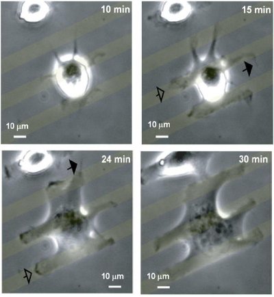 |
Figure 1: Images are single frames from a timelapse of a MEF spreading on 10 µm fibronectin stripes. |
Within the field of stimuli-responsive surfaces, Dr. Mendes' research group has developed6 electro-active surfaces that have been successfully employed to switch on functionalities in situ, offering an unprecedented ability to manipulate the interactions of proteins with surfaces. A new class of switchable biological surfaces - surface-confined electro-switchable peptides - have been also generated7 that have the capacity to regulate biomolecular interactions in response to an applied electrical potential.
This system is based upon the conformational switching of positively charged oligolysine peptides that are tethered to a gold surface, such that bioactive molecular moieties - biotin - incorporated on the end of the oligolysines can be exposed (bio-active state) or concealed (bio-inactive state) on demand (Figure 2), as a function of surface potential. The oligolysine peptides exhibit protonated amino side chains at pH =7, providing the basis for the switching "On" and "Off" of the biological activity on the surface.
For instance, upon application of a negative potential, the positively charged molecular system experiences an electrostatic attraction to the surface, leading to a mechanical molecular motion that shields the bioactive moiety (Figure 2). In order to test the viability of the proposed switching mechanism we selected an end functionalised biotinylated peptide as the biorecognition motif, four lysine residues as the switching unit and a terminal cysteine for self-assembled monolayer (SAM) formation on gold surfaces - biotin-Lys-Lys-Lys-Lys-Cys (Biotin-KKKKC).
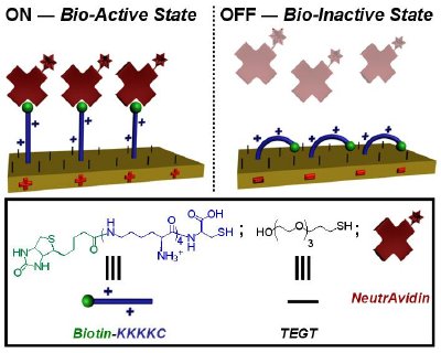 |
Figure 2. Schematic representation of the switching of mixed TEGT-biotinylated peptide SAMs between a bio-active and bio-inactive state. Depending on the electrical potential applied, the peptide can expose or conceal the biotin site and regulate its binding to NeutrAvidin. |
Apart from ensuring sufficient spatial freedom for synergistic molecular reorientation of the surface-bound biotinylated peptide, the short oligo(ethylene glycol) groups prevent nonspecific interaction from the proteins. As a control, a two-component, mixed SAM was also prepared using TEGT and a peptide without the biotin moiety - KKKKC. The dynamics of switching the biological properties was studied by observing the binding events between biotin and fluorescently labeled NeutrAvidin.
Fluorescence microscope images and SPR spectral data clearly revealed binding at + 0.3 V, reduced binding at - 0.4 V, and intermediate binding in the at open circuit (OC) conditions (no applied potential). Depending on the electrical potential applied to the mixed SAMs, bioactive molecules incorporated onto the SAM can be fully exposed for binding (+ 0.3 V, bio-active state) or concealed (- 0.4 V, bio-inactive state) to the extent that binding affinity can be reduced to over 90% of its bio-active state (Figure 3). Furthermore, reversibility studies by SPR also demonstrated that the developed switchable surface allows reversible control of biomolecular interactions.
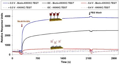 |
Figure 3. SPR sensorgram traces showing the binding of NeutrAvidin to the Biotin-KKKKC:TEGT mixed SAMs and KKKKC:TEGT mixed SAMs under OC conditions and an applied positive (+ 0.3 V) and negative (- 0.4 V) potential. |
References
1. C. M. Niemeyer, C. A. Mirkin, Wiley-VCH Verlag GmbH & Co. KGaA, Weinheim, 2004.2. P. M. Mendes, C. L. Yeung, J. A. Preece, Nanoscale Research Letters 2007, 2, 373.
3. P. M. Mendes, Chemical Society Reviews 2008, 37, 2512.
4. S. A. Johnston, J. P. Bramble, C. L. Yeung, P. M. Mendes, L. M. Machesky, BMC Cell Biology 2008, 9, 65.
5. C. Costello, J.-U. Kreft, C. M. Thomas, P. M. Mendes, Nanoproteomics: Methods and Protocols, S. Toms and R. Weil, eds., in Springer Protocols. Methods in Molecular Biology, Humana Press. In Press.
6. P. M. Mendes, K. L. Christman, P. Parthasarathy, E. Schopf, J. Ouyang, Y. Yang, J. A. Preece, H. D. Maynard, Y. Chen, J. F. Stoddart, Bioconjugate Chemistry 2007, 18, 1919.
7. C. L. Yeung, P. Iqbal, M. Allan, M. Lashkor, J. A. Preece, P. M. Mendes, Advanced Functional Materials, 2010, 20, 2657.
Copyright AZoNano.com, Dr. Paula Mendes (University of Birmingham)
Tidak ada komentar:
Posting Komentar